Optical imaging – Bioluminescence and fluorescence
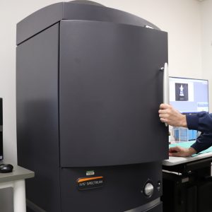
Perkin-Elmer IVIS Spectrum
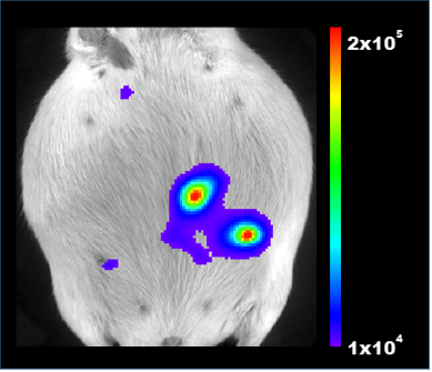
 This versatile, advanced in vivo bioluminescent and fluorescent pre-clinical imaging system combines high-throughput and full tomographic optical imaging in one platform. The light-tight imaging chamber has a heated stage and integrated gas anesthesia and allows for non-invasive longitudinal monitoring of disease progression, cell trafficking and gene expression patterns in living animals. An optimized set of high efficiency filters and advanced spectral unmixing algorithms allow spectral scanning over blue to near infrared wavelengths with visualization of multiple reporters (bioluminescent and/or fluorescent) in the same animal. 3D diffuse tomography for both bioluminescent and fluorescent optical signals can be quantified to determine source location and concentration, and analyzed to provide anatomical context following co-registration with the digital mouse atlas or other imaging modalities like µCT, MPI or MRI).
Technical Specifications:
- High-sensitivity in vivo imaging of fluorescence and bioluminescence
- High throughput (5 mice) with 23 cm field of view
- High resolution (to 20 microns) with 3.9 cm field of view
- Twenty eight high efficiency filters spanning 430 – 850 nm
- Supports spectral unmixing applications
- Ideal for distinguishing multiple bioluminescent and fluorescent reporters
- 3D diffuse tomographic reconstruction for both fluorescence and bioluminescence
- Import and automatically co-register CT or MRI images
- Isoflurane vaporizer