3D Histology (Emit Xerra)
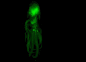
Use this system to automatically section and image samples ranging from organs to whole animals. Samples are embedded in OCT and frozen overnight, then placed in the chamber of the Xerra for sectioning. Images are then reconstructed into a 3D file for further analysis. Ideal for specimens with fluorescent tracers or particles, or genetically-encoded expression of fluorescence in structures of interest. There is the capacity to recover some sections onto a slide for staining.
Technical specifications:
White light and fluorescence imaging
Narrow band emission and excitation
Excitation 470-780 nm, emission 500-850 nm
Field of view up from 8×5 cm to 24×14 cm
In-plane resolution 30 µm x 100 µm, slice thickness 10 µm – 50 µm
Supports manual section collection
3D reconstruction software and data viewing