Magnetic Particle Imaging
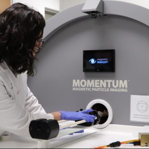
Magnetic Insight Momentum

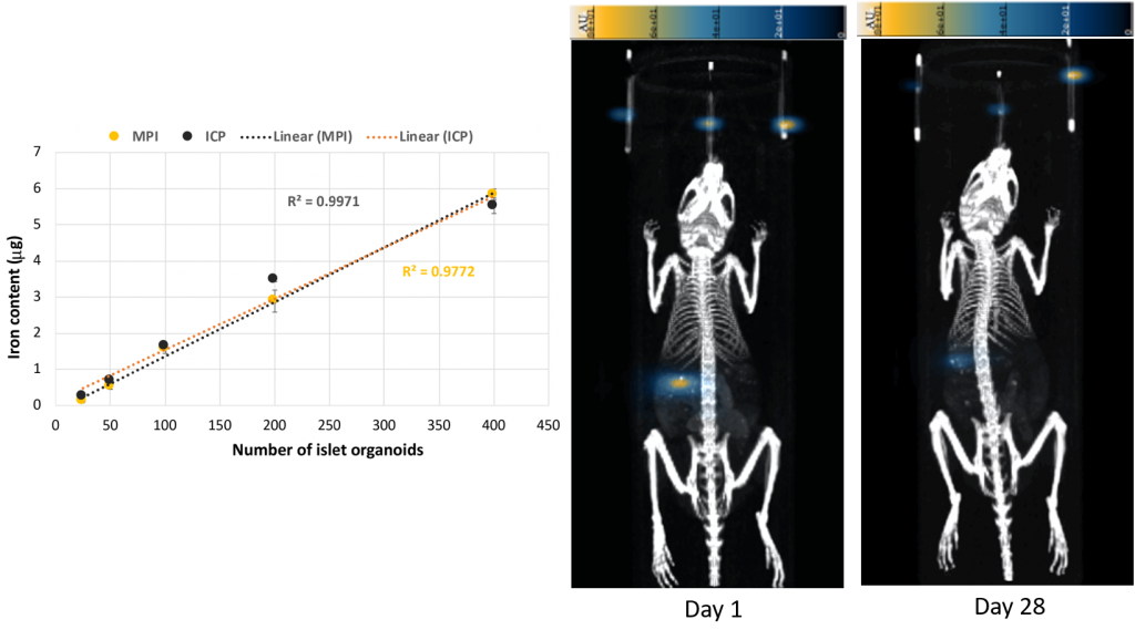
MPI sensitively and specifically detects superparamagnetic iron oxide particles in mice and can be applied to tracking immune cells, cancer metastasis, and targeted nanoparticles, for example. Particles may be injected intravenously or be used to label cells in vitro before they are administered to the mice. The images are positive contrast, with little to no background. 2D or 3D images can be acquired, and are easily co-registered with μCT through the use of a shuttle with dual CT/MPI fiducial markers. A relaxometry module is included for assessing new nanoparticles as MPI agents. Resolution and image quality are highly dependent on the particle used, so new agents are a strong research area.

Technical Specifications:
- Field free line field shape with mechanical field rotation
- 0-5.7 T/m (variable) field strength
- Water-cooled iron core electromagnet
- X gradient coil set (two individual main coils) magnet configuration
- Field of view: 6 cm x 6 cm x 12 cm
- RF transmit strength: >15 mT peak in (X,Z)
- 2D projection and 3D tomographic imaging sequences
- Animal bed with fiducials for µCT co-registration
- RELAX module included for particle characterization
- Isoflurane vaporizer